Torsades de pointes ekg examples - wikidoc is a high-quality image in the Cleve collection, available at 3249 × 2009 pixels resolution — ideal for both digital and print use.
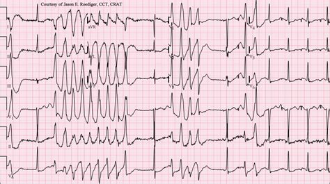
Learn to identify Torsades de Pointes on an ECG with this comprehensive guide. We cover critical diagnostic criteria, underlying causes like QT prolongation, and effective clinical management strategies. Understand the specific morphology of this life-threatening polymorphic ventricular tachycardia and improve your rhythm interpretation skills to ensure rapid intervention and better patient outcomes in emergency cardiac care.
Image Details
| Title | Torsades de pointes ekg examples - wikidoc |
|---|---|
| Dimensions | 3249 × 2009 px |
| Category | Cleve |
| Published | October 25, 2024 |
| Author | Zeus |
| Downloads | 2,204 |
| Views | 542 |
Frequently Asked Questions
This image has a resolution of 3249 × 2009 pixels. It is suitable for high-quality printing, digital presentations, and web use without losing clarity.
This image is part of the Cleve collection. You can browse more images in this category to find similar content.
Click the Download button above the image to save it directly to your device. The image is provided in its original resolution of 3249 × 2009 px.
Yes! Scroll down to the More Images section below to explore related Cleve images. You can also visit the full article for more context and a complete image gallery.
Read full article: Torsades De Pointes Ecg