Sphenoid bone: Anatomy, function and development | Kenhub is a high-quality image in the Rp collection, available at 1400 × 1400 pixels resolution — ideal for both digital and print use.
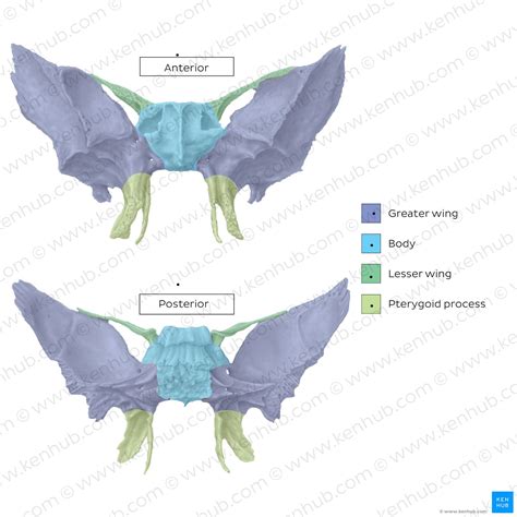
Explore the anatomy and clinical significance of the Sphenoid Sella Turcica. Learn about this saddle-shaped bone depression, its vital role in protecting the pituitary gland, and its importance in diagnosing endocrine disorders, skull base imaging, and neurosurgical procedures. Understand how variations in this sphenoid bone structure impact diagnostic accuracy and patient health outcomes in modern clinical practice.
Image Details
| Title | Sphenoid bone: Anatomy, function and development | Kenhub |
|---|---|
| Dimensions | 1400 × 1400 px |
| Category | Rp |
| Published | November 15, 2025 |
| Author | Zeus |
| Downloads | 1,364 |
| Views | 835 |
Frequently Asked Questions
This image has a resolution of 1400 × 1400 pixels. It is suitable for high-quality printing, digital presentations, and web use without losing clarity.
This image is part of the Rp collection. You can browse more images in this category to find similar content.
Click the Download button above the image to save it directly to your device. The image is provided in its original resolution of 1400 × 1400 px.
Yes! Scroll down to the More Images section below to explore related Rp images. You can also visit the full article for more context and a complete image gallery.
Read full article: Sphenoid Sella Turcica