Posterior Cranial Fossa is a high-quality image in the Rp collection, available at 1350 × 1800 pixels resolution — ideal for both digital and print use.
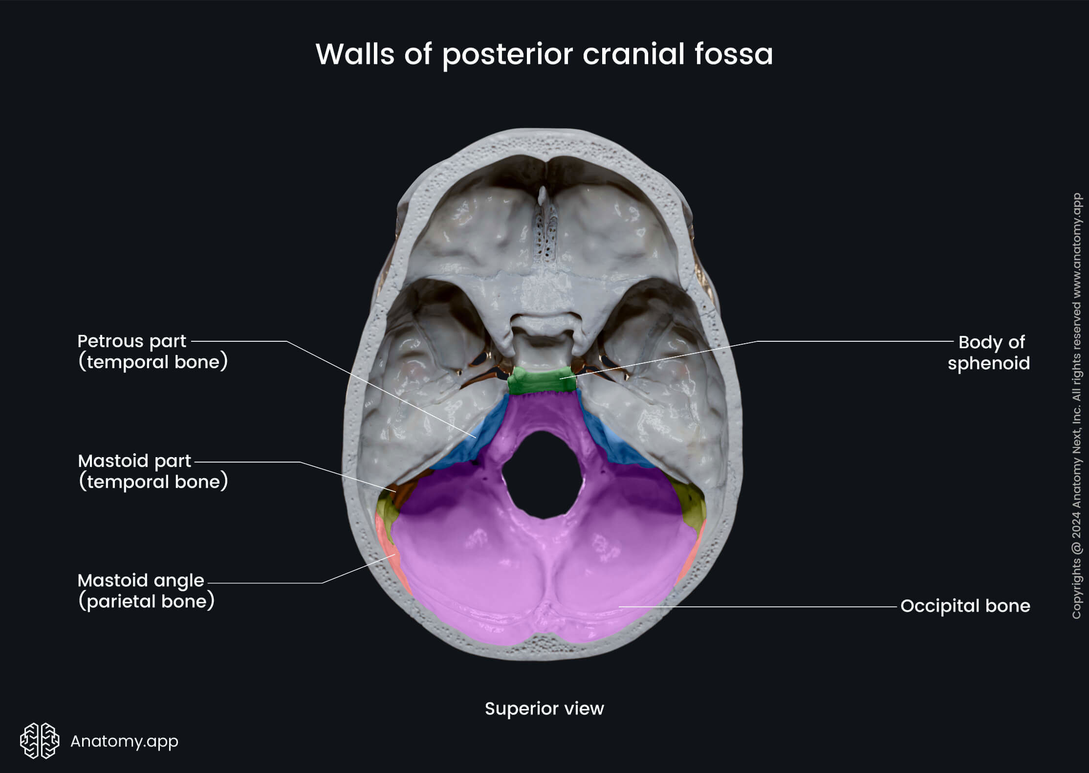
Explore the anatomy and clinical significance of the skull posterior fossa. This guide details essential structures, including the cerebellum and brainstem, and examines common pathologies such as tumors and Chiari malformations. Learn how advanced diagnostic imaging helps clinicians identify cranial abnormalities within the posterior cranial compartment to improve patient outcomes and support surgical planning.
Image Details
| Title | Posterior Cranial Fossa |
|---|---|
| Dimensions | 1350 × 1800 px |
| Category | Rp |
| Published | October 13, 2024 |
| Author | Zeus |
| Downloads | 1,199 |
| Views | 1,830 |
Frequently Asked Questions
This image has a resolution of 1350 × 1800 pixels. It is suitable for high-quality printing, digital presentations, and web use without losing clarity.
This image is part of the Rp collection. You can browse more images in this category to find similar content.
Click the Download button above the image to save it directly to your device. The image is provided in its original resolution of 1350 × 1800 px.
Yes! Scroll down to the More Images section below to explore related Rp images. You can also visit the full article for more context and a complete image gallery.
Read full article: Skull Posterior Fossa