X Ray Shoulder Anatomy is a high-quality image in the Cleve collection, available at 1637 × 1215 pixels resolution — ideal for both digital and print use.
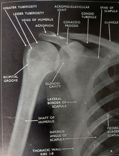
Learn what to expect during a shoulder X-ray. This guide explains how this diagnostic imaging procedure identifies fractures, dislocations, or arthritis. Discover how medical professionals use these X-ray images to assess joint health, evaluate bone structures, and determine the best treatment plan for your shoulder pain or injury. Optimize your preparation for an accurate clinical diagnosis today.
Image Details
| Title | X Ray Shoulder Anatomy |
|---|---|
| Dimensions | 1637 × 1215 px |
| Category | Cleve |
| Published | February 9, 2025 |
| Author | Zeus |
| Downloads | 596 |
| Views | 921 |
Frequently Asked Questions
This image has a resolution of 1637 × 1215 pixels. It is suitable for high-quality printing, digital presentations, and web use without losing clarity.
This image is part of the Cleve collection. You can browse more images in this category to find similar content.
Click the Download button above the image to save it directly to your device. The image is provided in its original resolution of 1637 × 1215 px.
Yes! Scroll down to the More Images section below to explore related Cleve images. You can also visit the full article for more context and a complete image gallery.
Read full article: Shoulder X Ray