Brain Parenchymal Signal Abnormalities Associated with Developmental Venous Anomalies: Detailed ... is a high-quality image in the Cleve collection, available at 1800 × 1045 pixels resolution — ideal for both digital and print use.
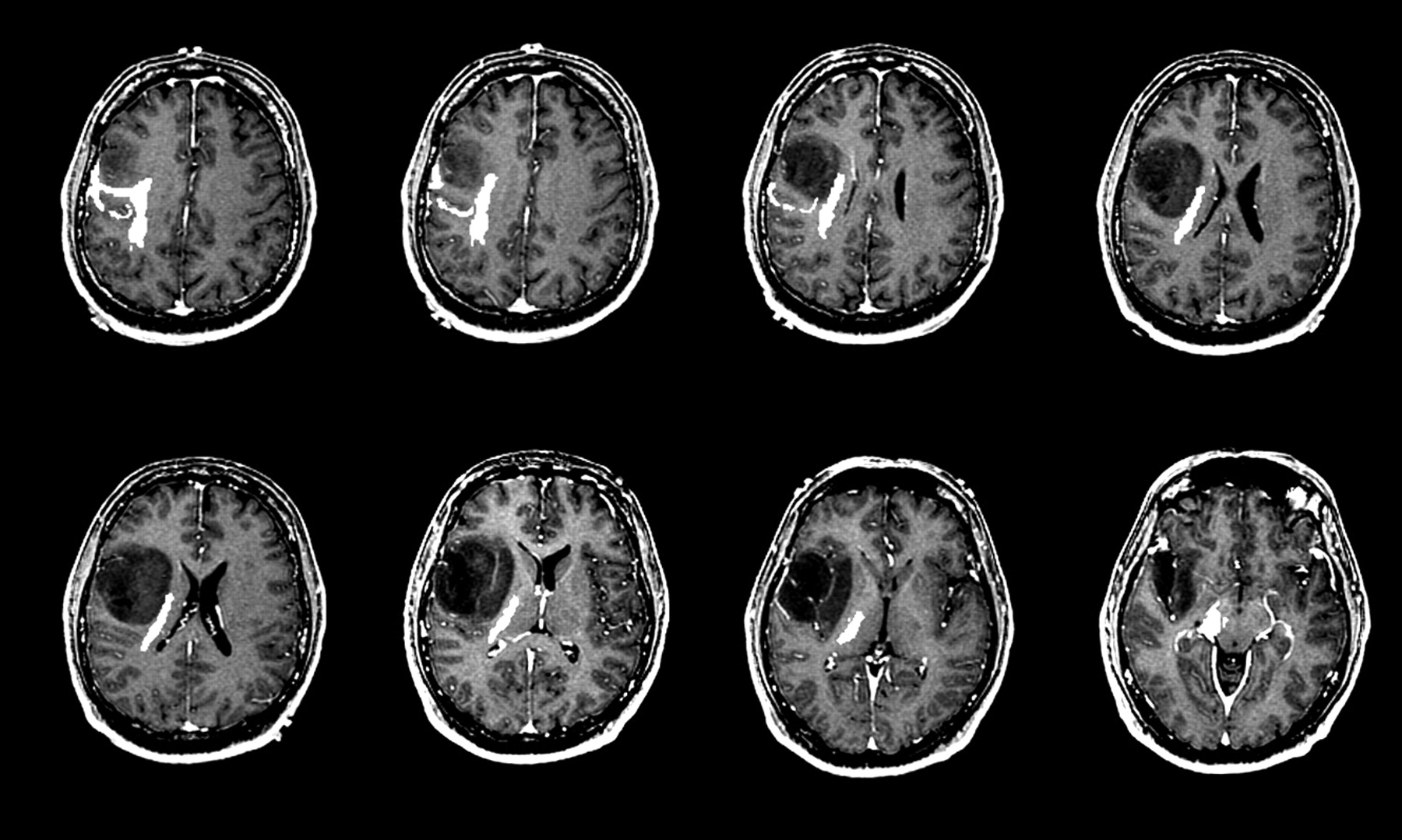
Understand the implications of parenchymal volume loss on brain health. This guide explains how reduced gray and white matter relates to neurodegenerative conditions like Alzheimer’s, highlighting key diagnostic imaging, common causes, and current clinical research. Learn why monitoring brain atrophy is essential for early detection and effective cognitive impairment management in aging patients.
Image Details
| Title | Brain Parenchymal Signal Abnormalities Associated with Developmental Venous Anomalies: Detailed ... |
|---|---|
| Dimensions | 1800 × 1045 px |
| Category | Cleve |
| Published | April 10, 2025 |
| Author | Zeus |
| Downloads | 462 |
| Views | 917 |
Frequently Asked Questions
This image has a resolution of 1800 × 1045 pixels. It is suitable for high-quality printing, digital presentations, and web use without losing clarity.
This image is part of the Cleve collection. You can browse more images in this category to find similar content.
Click the Download button above the image to save it directly to your device. The image is provided in its original resolution of 1800 × 1045 px.
Yes! Scroll down to the More Images section below to explore related Cleve images. You can also visit the full article for more context and a complete image gallery.
Read full article: Parenchymal Volume Loss