Ecg Leads is a high-quality image in the Ghc collection, available at 3361 × 1948 pixels resolution — ideal for both digital and print use.
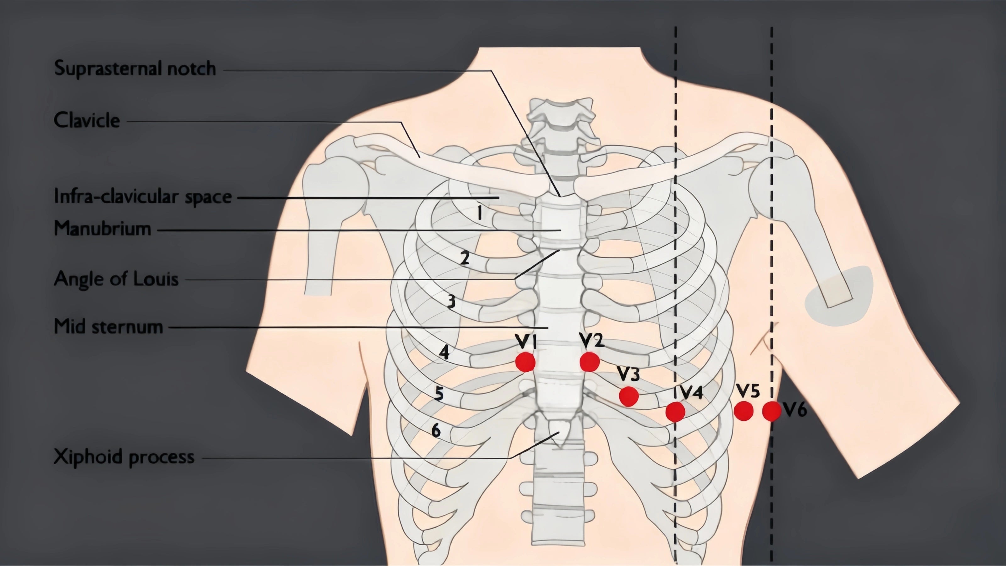
Master the interpretation of lateral leads of ECG to identify myocardial ischemia. Learn how leads I, aVL, V5, and V6 provide critical insights into the heart's lateral wall, aiding accurate cardiac diagnosis. Enhance your clinical skills with our comprehensive guide on lead placement, diagnostic waveforms, and recognizing ST-segment changes for better patient outcomes in emergency and routine cardiology.
Image Details
| Title | Ecg Leads |
|---|---|
| Dimensions | 3361 × 1948 px |
| Category | Ghc |
| Published | November 22, 2024 |
| Author | Zeus |
| Downloads | 549 |
| Views | 1,661 |
Frequently Asked Questions
This image has a resolution of 3361 × 1948 pixels. It is suitable for high-quality printing, digital presentations, and web use without losing clarity.
This image is part of the Ghc collection. You can browse more images in this category to find similar content.
Click the Download button above the image to save it directly to your device. The image is provided in its original resolution of 3361 × 1948 px.
Yes! Scroll down to the More Images section below to explore related Ghc images. You can also visit the full article for more context and a complete image gallery.
Read full article: Lateral Leads Of Ecg