Triquetral fracture - X Rays Case Studies - CTisus CT Scanning is a high-quality image in the Rp collection, available at 1080 × 1297 pixels resolution — ideal for both digital and print use.
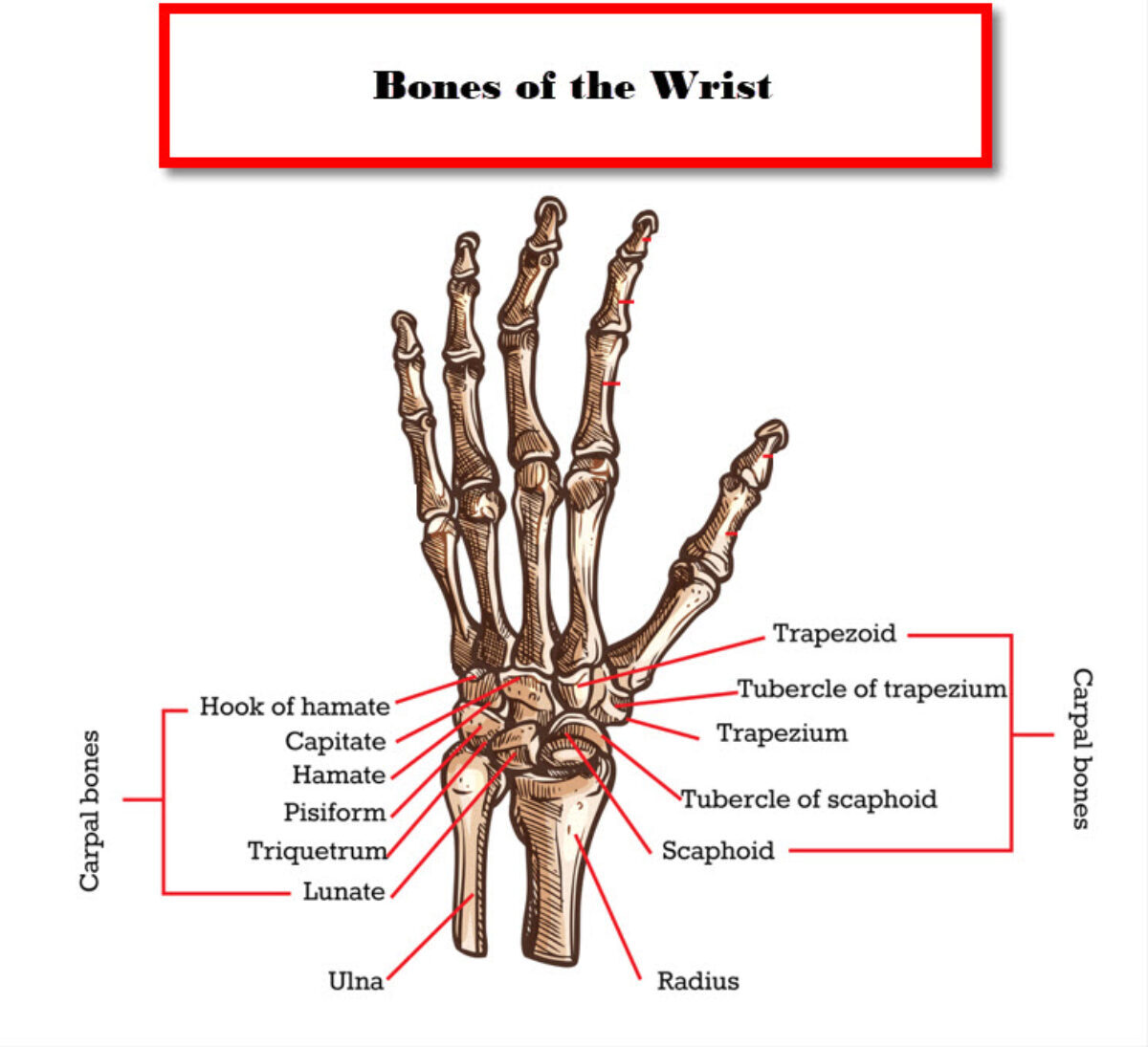
Learn the essential facts about a fracture triquetral bone, including common causes, symptoms, and effective treatment options. Discover how to recognize wrist pain and navigate recovery protocols for this carpal injury. Our comprehensive guide helps you understand diagnosis, immobilization techniques, and rehabilitation to ensure proper healing and restore full range of motion to your injured wrist.
Image Details
| Title | Triquetral fracture - X Rays Case Studies - CTisus CT Scanning |
|---|---|
| Dimensions | 1080 × 1297 px |
| Category | Rp |
| Published | April 2, 2025 |
| Author | Zeus |
| Downloads | 2,265 |
| Views | 1,886 |
Frequently Asked Questions
This image has a resolution of 1080 × 1297 pixels. It is suitable for high-quality printing, digital presentations, and web use without losing clarity.
This image is part of the Rp collection. You can browse more images in this category to find similar content.
Click the Download button above the image to save it directly to your device. The image is provided in its original resolution of 1080 × 1297 px.
Yes! Scroll down to the More Images section below to explore related Rp images. You can also visit the full article for more context and a complete image gallery.
Read full article: Fracture Triquetral Bone