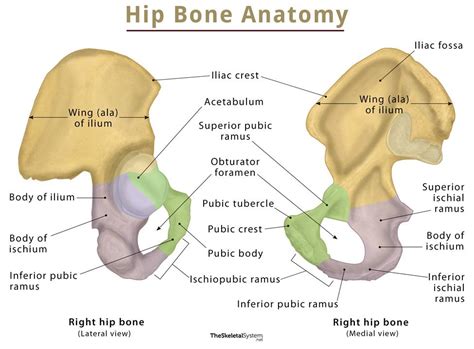
Coxal Bone Anatomy is a high-quality image in the Cleve collection, available at 1299 × 1390 pixels resolution — ideal for both digital and print use.
Discover the anatomy and functions of the coxal hip bone. Learn how this vital pelvic structure, comprising the ilium, ischium, and pubis, supports skeletal stability, facilitates movement, and protects internal organs. Explore detailed insights into hip joint mechanics and the role of the innominate bone in human anatomy to better understand your body’s complex musculoskeletal framework.
Image Details
| Title | Coxal Bone Anatomy |
|---|---|
| Dimensions | 1299 × 1390 px |
| Category | Cleve |
| Published | July 27, 2025 |
| Author | Zeus |
| Downloads | 2,463 |
| Views | 205 |
Frequently Asked Questions
This image has a resolution of 1299 × 1390 pixels. It is suitable for high-quality printing, digital presentations, and web use without losing clarity.
This image is part of the Cleve collection. You can browse more images in this category to find similar content.
Click the Download button above the image to save it directly to your device. The image is provided in its original resolution of 1299 × 1390 px.
Yes! Scroll down to the More Images section below to explore related Cleve images. You can also visit the full article for more context and a complete image gallery.
Read full article: Coxal Hip Bone