Brugada Ecg – Brugada 心臓 , LE SYNDROME DE BRUGADA – APTR is a high-quality image in the Ghc collection, available at 3000 × 1687 pixels resolution — ideal for both digital and print use.
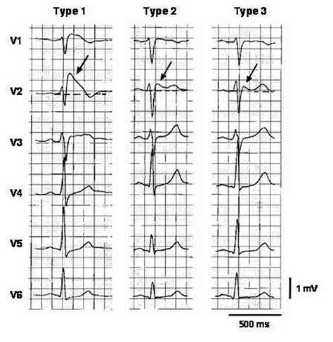
Learn how to identify Brugada Syndrome ECG patterns to improve patient diagnosis and outcomes. Our guide covers essential diagnostic criteria, key cardiac markers, and the distinct coved or saddleback ST-segment elevations. Understand the clinical significance of these rhythm abnormalities and how early detection of this inherited arrhythmia disorder can help prevent sudden cardiac arrest and manage long-term risks.
Image Details
| Title | Brugada Ecg – Brugada 心臓 , LE SYNDROME DE BRUGADA – APTR |
|---|---|
| Dimensions | 3000 × 1687 px |
| Category | Ghc |
| Published | February 8, 2026 |
| Author | Zeus |
| Downloads | 174 |
| Views | 955 |
Frequently Asked Questions
This image has a resolution of 3000 × 1687 pixels. It is suitable for high-quality printing, digital presentations, and web use without losing clarity.
This image is part of the Ghc collection. You can browse more images in this category to find similar content.
Click the Download button above the image to save it directly to your device. The image is provided in its original resolution of 3000 × 1687 px.
Yes! Scroll down to the More Images section below to explore related Ghc images. You can also visit the full article for more context and a complete image gallery.
Read full article: Brugada Syndrome Ecg