RADIOGRAPHIC ANATOMY OF KNEE JOINT AND ITS RADIOGRAPHIC VIEWS.pptx is a high-quality image in the Cleve collection, available at 2048 × 1152 pixels resolution — ideal for both digital and print use.
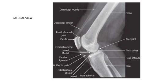
Master the complex anatomy for knee function with this comprehensive guide. We break down essential components, including the femur, tibia, patella, ligaments, and cartilage. Understand how these structures work together to facilitate movement, prevent common injuries, and maintain joint health. Enhance your knowledge of orthopedic mechanics and human biology with this clear, expert overview of the knee joint.
Image Details
| Title | RADIOGRAPHIC ANATOMY OF KNEE JOINT AND ITS RADIOGRAPHIC VIEWS.pptx |
|---|---|
| Dimensions | 2048 × 1152 px |
| Category | Cleve |
| Published | September 29, 2025 |
| Author | Zeus |
| Downloads | 1,532 |
| Views | 2,394 |
Frequently Asked Questions
This image has a resolution of 2048 × 1152 pixels. It is suitable for high-quality printing, digital presentations, and web use without losing clarity.
This image is part of the Cleve collection. You can browse more images in this category to find similar content.
Click the Download button above the image to save it directly to your device. The image is provided in its original resolution of 2048 × 1152 px.
Yes! Scroll down to the More Images section below to explore related Cleve images. You can also visit the full article for more context and a complete image gallery.
Read full article: Anatomy For Knee